Orka
From .org
- Joined
- Sep 4, 2025
- Posts
- 147
- Reputation
- 392
The Radiology behind Facial Development & Bodily Growth Indicators
I've seen at least 50 questions asking if somebody's growth plates are still open or not, or to estimate their bone age,so to make it easy, here's how you can answer your own questions!
Table Of Contents (easiest to hardest)
1. What are growth plates & how to determine their status
2. Facial Growth Centers & Sutures
3. How to estimate bone age
1. What are growth plates & how to determine their status
A growth plate (or an Epiphyseal plate) is a region of cartilage near the ends of long bones in children and adolescents where new bone tissue is created, causing the bone to grow in length and width.
This process continues until adolescence, when the growth plates harden into solid bone, a process called "closing" or “fusing”.
Male growth plates tend to fuse at around 16-19, once your plates have fused/closed, they will not grow any longer or wider.
Now, to find out if your growth plates are fused or not, we’re going to look at your wrist & knees for your height, and at your clavicles for your frame.

Above I've added 3 figures, from open ( A ) to fully fused ( C ), those are your growth plates, if theres a visible line like in B, they’re still open, but closer to fusion. ( You will typically see this in X-rays of teenagers). Whereas if you don’t see any lines at all, it means the cartilage has hardened into solid bone, ending any growth potential.

Once again, refer to the graphic above.
If there’s a visible gap or a full line, your growth plates are open, if there’s no visible line, they’re closed.
2. Facial Growth Centers & Sutures
The most important sutures when it comes to facial development are your Midpalatal, Frontomaxillary, Zygomatomaxillary & Pterygopalatine Sutures, but in reality your sutures aren't a growth center, meaning that scanning them and determining their status is for the most part useless, except for the midpalatal suture (one that is a little bit important!)
i. Midpalatal Suture

Here are graphics (I) and CBCT Scans (II) of your midpalatal suture.
Figure A is a young child, 10-12 years old, then onto Figure B where the child is 11-13, C where they’re 13-15, D representing a 15-19 year old, and E representing a fully fused midpalatal suture of a fully grown person, usually 19-20+ years old.
If you still see a line on your scan, your suture is not fused. If you do not see any line on your scan, it’s fused.
Your midpalatal suture begins fusing from the back, so the line you do see will get shorter & shorter until eventually it matures and fuses completely.
Clinically speaking the only significant thing you need to take away from your scans are if your midpalatal suture is fused or not, around the ages of 15-19 you’ll likely have this scan done by your ortho if you’re considering getting a palate expander of any kind.
If it is fused, then you’ll likely be assigned a SARME, SARPE or SAMARPE for your expansion ( Note that this will not mean your zygos become wider, or any extreme external aesthetic benefit, your palate will get wider. ) whereas if it’s not fused, you’ll typically be assigned a regular mse / expander.
ii. Aesthetic Relevance of Sutures.
Before joining this forum I was super active in many looksmaxing discord servers, where I noticed people believing Sutures worked similarly to growth plates.
I’m not saying you believe this, I’m adding this section to clear up misconceptions.
Your sutures are not growth centers, they do not grow & just because your sutures are open doesn’t mean they’ll grow.
The status of your sutures simply shows if the area will respond to growth or not.
If your sutures are closed, no you won’t grow anymore, but just because they’re open doesn’t mean they’ll grow, so even if you notice all your sutures are open, your growth still might be over.
iii. Nasal Septal Cartilage
As opposed to Sutures, The Nasal Septal Cartilage is a growth center, meaning its made of the same Hyaline cartilage that your growth plates are made of.

This area is mainly responsible for growing your midface forward, or downwards (oh no!).
Now, the moment this cartilage stops growing, the development of your midface stops aswell.
This is cartilage, meaning it doesn’t show up xrays, or CT/CBCT Scans, the only place you can actually see it is on MRI’s, but there’s literally no way you can bullshit your way into getting an MRI, especially for something like this.
Now the typical method to determine this is actually through lefthand xrays, hyaline cartilage ossifying is systematic, which is why you can check your knee or your wrist to determine your growth plate status, and not have to check both or prioritize one of them.
It’s as simple as this, if the growth plates in your wrist or knee are fused (see Chapter 1 of this guide to determine that), then your nasal septal cartilage has also ossified and completed its work. (Most of the time, after one growth site in your body has ossified, the others follow suit within the next few months, not enough time to get any significant growth anyways).
3. How to estimate your bone age. (Better than ChatGPT)
In my personal experience experimenting with this method, my estimates have beaten chatgpt 3/3 times.
I’m not an experienced radiologist, and asking chatgpt is the only other option for users to estimate their bone age without going to a radiologist (This isn’t to replace a radiologist’s rating, this is for an at-home analysis)
The method we’re going to be using is the Tanner-Whitehouse 3, Radius-Ulna-Short Bones method. For simplicity’s sake I’ll refer to it as the TW3-RUS method from now on.
The typical margin of error using this method is 6-9 months, although during puberty this can differ up to over a year since people can have unpredictable and uneven growth spurts.
TW3-RUS is the most refined and as of today the best method for estimating bone age.
To continue with this method & estimate your own bone age, you’ll need an xray of your non dominant hand, including your wrist. We’re going to be evaluating a total of 13 bones.
I’ve worked my ass off to create the following reference sheets, so here’s what you’re going to do with them.
I added references for stages A (Completely unfused), E-F (Partial fusion, basically late stages of puberty), and I (Complete Fusion)
You will have to eyeball this now using your own xrays, I’ve given three examples and some wiggle-room for letters using a reference bar (green-red) below the image, write down your estimated guesses in a text file until you finish all 13 bones.
Don’t doubt yourself, eyeballing isn’t perfect but it still works well, just choose & continue.
Ex.
“My Radius looks more fused than the image from E-F, but its not as fused as I, it’s closer to the 2nd picture though, so I’ll rate it G” And continue
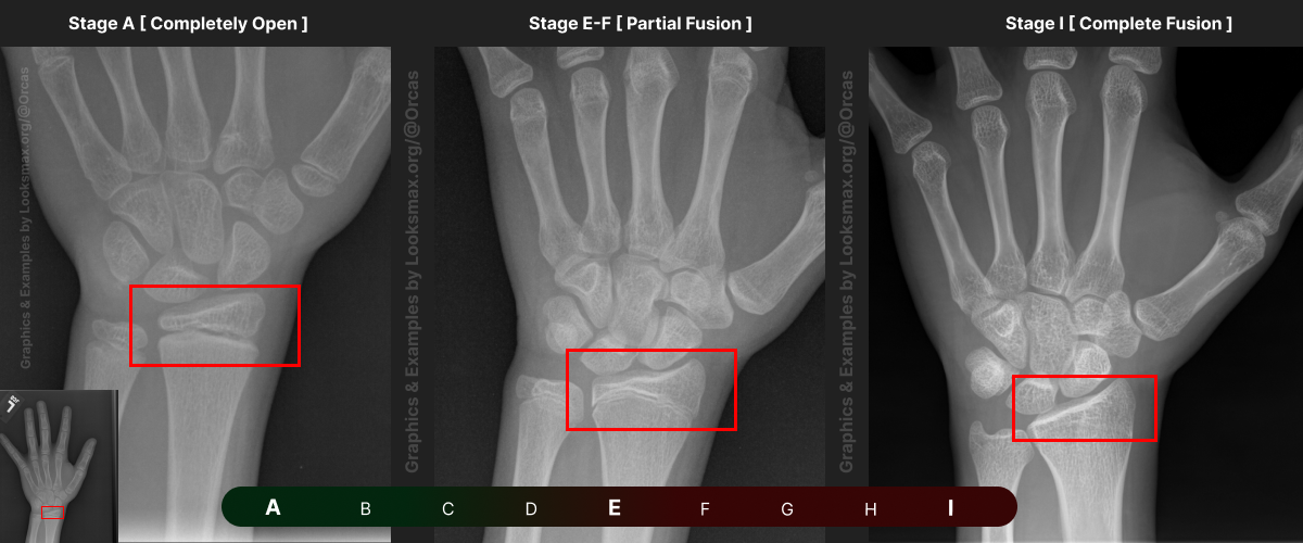
Bonetype: Radius (distal end)
.png")
Check & use the chart below to figure out where you are. Then write your result on a piece of paper or a text file.
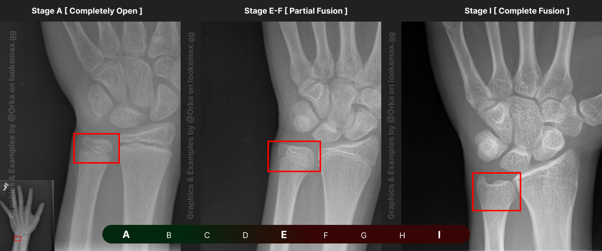
Bonetype: Ulna (distal end)
.png")
Check & use the chart below to figure out where you are. Then write your result on a piece of paper or a text file.
Bonetype: 1st Metacarpal (thumb)
.png")
Check & use the chart below to figure out where you are. Then write your result on a piece of paper or a text file.
Bonetype: 3rd Metacarpal (middle finger)
.png")
Check & use the chart below to figure out where you are. Then write your result on a piece of paper or a text file.
Bonetype: 5th Metacarpal (pinky)
.png")
Check & use the chart below to figure out where you are. Then write your result on a piece of paper or a text file.
Bonetype: 1st Proximal Phalanx

Check & use the chart below to figure out where you are. Then write your result on a piece of paper or a text file.
Bonetype: 3rd Proximal Phalanx

Check & use the chart below to figure out where you are. Then write your result on a piece of paper or a text file.
Bonetype: 5th Proximal Phalanx

Check & use the chart below to figure out where you are. Then write your result on a piece of paper or a text file.
Bonetype: 3rd Middle Phalanx

Check & use the chart below to figure out where you are. Then write your result on a piece of paper or a text file.
Bonetype: 5th Middle Phalanx

Check & use the chart below to figure out where you are. Then write your result on a piece of paper or a text file.
Bonetype: 1st Distal Phalanx (thumb)
.png")
Check & use the chart below to figure out where you are. Then write your result on a piece of paper or a text file.
Bonetype: 3rd Distal Phalanx (middle finger)
.png")
Check & use the chart below to figure out where you are. Then write your result on a piece of paper or a text file.
Bonetype: 5th Distal Phalanx (pinky)
.png")
Check & use the chart below to figure out where you are. Then write your result on a piece of paper or a text file.
There you go, it wasn’t that hard was it?
Now that you’ve got your letters written down, it should look something like this:
Radius = H, Ulna = G
1st MC = I, 3rd MC = I, 5th MC = H
1st PProx = I, 3rd PProx = I, 5th PProx = H
3rd PMid = H, 5th PMid = G
1st PDist = I, 3rd PDist = H, 5th PDist = H
( You should have labeled them ideally, and the letters will vary )
Now convert these letters into numerical value ( TW3-RUS Score )

For each letter you should assign points per bone, then calculate the total by combining all of the points
There’s your bone age within a 6-9 month range. You can consider adding another slight margin of error since you’re not an experienced radiologist & don’t have experience reading these.
Bone age = chronological age = normal growth tempo ( You’ll grow normally )
Bone age < chronological age = delayed skeletal maturation ( You’ll grow more )
Bone age > chronological age = advanced skeletal maturation ( You’ll grow less )
If you want to practice & get better at this, I’ve left a link to a 10gb dataset of left hand xrays (with the correct result bone age assigned) as a link below
I hope this helped, and I hope I didn’t annihilate your dreams of growing taller!
Tags: BigBallsLarry
 Kaligula567
 Steve Rogers ( I don't know anybody else on here tbh, not very active)
I've seen at least 50 questions asking if somebody's growth plates are still open or not, or to estimate their bone age,so to make it easy, here's how you can answer your own questions!
Table Of Contents (easiest to hardest)
1. What are growth plates & how to determine their status
- What are growth plates?
- Analysis of your wrist’s growth plate
- Analysis of your knee’s growth plate
2. Facial Growth Centers & Sutures
- Midpalatal Suture
- Aesthetic Relevance of Sutures in your face
- Nasal Septal Cartilage
3. How to estimate bone age
- An Introduction to TW3-RUS
- Where to look & how to determine sizes [ Custom reference sheets ]
- Process walkthrough & conclusion
1. What are growth plates & how to determine their status
A growth plate (or an Epiphyseal plate) is a region of cartilage near the ends of long bones in children and adolescents where new bone tissue is created, causing the bone to grow in length and width.
This process continues until adolescence, when the growth plates harden into solid bone, a process called "closing" or “fusing”.
Male growth plates tend to fuse at around 16-19, once your plates have fused/closed, they will not grow any longer or wider.
Now, to find out if your growth plates are fused or not, we’re going to look at your wrist & knees for your height, and at your clavicles for your frame.
Above I've added 3 figures, from open ( A ) to fully fused ( C ), those are your growth plates, if theres a visible line like in B, they’re still open, but closer to fusion. ( You will typically see this in X-rays of teenagers). Whereas if you don’t see any lines at all, it means the cartilage has hardened into solid bone, ending any growth potential.
Once again, refer to the graphic above.
If there’s a visible gap or a full line, your growth plates are open, if there’s no visible line, they’re closed.
2. Facial Growth Centers & Sutures
The most important sutures when it comes to facial development are your Midpalatal, Frontomaxillary, Zygomatomaxillary & Pterygopalatine Sutures, but in reality your sutures aren't a growth center, meaning that scanning them and determining their status is for the most part useless, except for the midpalatal suture (one that is a little bit important!)
i. Midpalatal Suture
Here are graphics (I) and CBCT Scans (II) of your midpalatal suture.
Figure A is a young child, 10-12 years old, then onto Figure B where the child is 11-13, C where they’re 13-15, D representing a 15-19 year old, and E representing a fully fused midpalatal suture of a fully grown person, usually 19-20+ years old.
If you still see a line on your scan, your suture is not fused. If you do not see any line on your scan, it’s fused.
Your midpalatal suture begins fusing from the back, so the line you do see will get shorter & shorter until eventually it matures and fuses completely.
Clinically speaking the only significant thing you need to take away from your scans are if your midpalatal suture is fused or not, around the ages of 15-19 you’ll likely have this scan done by your ortho if you’re considering getting a palate expander of any kind.
If it is fused, then you’ll likely be assigned a SARME, SARPE or SAMARPE for your expansion ( Note that this will not mean your zygos become wider, or any extreme external aesthetic benefit, your palate will get wider. ) whereas if it’s not fused, you’ll typically be assigned a regular mse / expander.
ii. Aesthetic Relevance of Sutures.
Before joining this forum I was super active in many looksmaxing discord servers, where I noticed people believing Sutures worked similarly to growth plates.
I’m not saying you believe this, I’m adding this section to clear up misconceptions.
Your sutures are not growth centers, they do not grow & just because your sutures are open doesn’t mean they’ll grow.
The status of your sutures simply shows if the area will respond to growth or not.
If your sutures are closed, no you won’t grow anymore, but just because they’re open doesn’t mean they’ll grow, so even if you notice all your sutures are open, your growth still might be over.
iii. Nasal Septal Cartilage
As opposed to Sutures, The Nasal Septal Cartilage is a growth center, meaning its made of the same Hyaline cartilage that your growth plates are made of.
This area is mainly responsible for growing your midface forward, or downwards (oh no!).
Now, the moment this cartilage stops growing, the development of your midface stops aswell.
This is cartilage, meaning it doesn’t show up xrays, or CT/CBCT Scans, the only place you can actually see it is on MRI’s, but there’s literally no way you can bullshit your way into getting an MRI, especially for something like this.
Now the typical method to determine this is actually through lefthand xrays, hyaline cartilage ossifying is systematic, which is why you can check your knee or your wrist to determine your growth plate status, and not have to check both or prioritize one of them.
It’s as simple as this, if the growth plates in your wrist or knee are fused (see Chapter 1 of this guide to determine that), then your nasal septal cartilage has also ossified and completed its work. (Most of the time, after one growth site in your body has ossified, the others follow suit within the next few months, not enough time to get any significant growth anyways).
3. How to estimate your bone age. (Better than ChatGPT)
In my personal experience experimenting with this method, my estimates have beaten chatgpt 3/3 times.
I’m not an experienced radiologist, and asking chatgpt is the only other option for users to estimate their bone age without going to a radiologist (This isn’t to replace a radiologist’s rating, this is for an at-home analysis)
The method we’re going to be using is the Tanner-Whitehouse 3, Radius-Ulna-Short Bones method. For simplicity’s sake I’ll refer to it as the TW3-RUS method from now on.
The typical margin of error using this method is 6-9 months, although during puberty this can differ up to over a year since people can have unpredictable and uneven growth spurts.
TW3-RUS is the most refined and as of today the best method for estimating bone age.
To continue with this method & estimate your own bone age, you’ll need an xray of your non dominant hand, including your wrist. We’re going to be evaluating a total of 13 bones.
I’ve worked my ass off to create the following reference sheets, so here’s what you’re going to do with them.
I added references for stages A (Completely unfused), E-F (Partial fusion, basically late stages of puberty), and I (Complete Fusion)
You will have to eyeball this now using your own xrays, I’ve given three examples and some wiggle-room for letters using a reference bar (green-red) below the image, write down your estimated guesses in a text file until you finish all 13 bones.
Don’t doubt yourself, eyeballing isn’t perfect but it still works well, just choose & continue.
Ex.
“My Radius looks more fused than the image from E-F, but its not as fused as I, it’s closer to the 2nd picture though, so I’ll rate it G” And continue
Bonetype: Radius (distal end)
Check & use the chart below to figure out where you are. Then write your result on a piece of paper or a text file.
Bonetype: Ulna (distal end)
Check & use the chart below to figure out where you are. Then write your result on a piece of paper or a text file.
Bonetype: 1st Metacarpal (thumb)
Check & use the chart below to figure out where you are. Then write your result on a piece of paper or a text file.
Bonetype: 3rd Metacarpal (middle finger)
Check & use the chart below to figure out where you are. Then write your result on a piece of paper or a text file.
Bonetype: 5th Metacarpal (pinky)
Check & use the chart below to figure out where you are. Then write your result on a piece of paper or a text file.
Bonetype: 1st Proximal Phalanx
Check & use the chart below to figure out where you are. Then write your result on a piece of paper or a text file.
Bonetype: 3rd Proximal Phalanx
Check & use the chart below to figure out where you are. Then write your result on a piece of paper or a text file.
Bonetype: 5th Proximal Phalanx
Check & use the chart below to figure out where you are. Then write your result on a piece of paper or a text file.
Bonetype: 3rd Middle Phalanx
Check & use the chart below to figure out where you are. Then write your result on a piece of paper or a text file.
Bonetype: 5th Middle Phalanx
Check & use the chart below to figure out where you are. Then write your result on a piece of paper or a text file.
Bonetype: 1st Distal Phalanx (thumb)
Check & use the chart below to figure out where you are. Then write your result on a piece of paper or a text file.
Bonetype: 3rd Distal Phalanx (middle finger)
Check & use the chart below to figure out where you are. Then write your result on a piece of paper or a text file.
Bonetype: 5th Distal Phalanx (pinky)
Check & use the chart below to figure out where you are. Then write your result on a piece of paper or a text file.
There you go, it wasn’t that hard was it?
Now that you’ve got your letters written down, it should look something like this:
Radius = H, Ulna = G
1st MC = I, 3rd MC = I, 5th MC = H
1st PProx = I, 3rd PProx = I, 5th PProx = H
3rd PMid = H, 5th PMid = G
1st PDist = I, 3rd PDist = H, 5th PDist = H
( You should have labeled them ideally, and the letters will vary )
Now convert these letters into numerical value ( TW3-RUS Score )
For each letter you should assign points per bone, then calculate the total by combining all of the points
| Total TW3-RUS Score | Estimated Bone age ( male ) | Estimated Bone age ( female ) |
| 400 | 9 Years 6 Months | 9 Years 3 Months |
| 425 | 10 Years 3 Months | 9 Years 9 Months |
| 450 | 11 Years 0 Months | 10 Years 0 Months |
| 475 | 11 Years 6 Months | 11 Years 0 Months |
| 500 | 12 Years 3 Months | 12 Years 0 Months |
| 525 | 12 Years 9 Months | 12 Years 6 Months |
| 550 | 13 Years 3 Months | 13 Years 0 Months |
| 575 | 13 Years 9 Months | 13 Years 6 Months |
| 600 | 14 Years 3 Months | 14 Years 0 Months |
| 625 | 14 Years 9 Months | 14 Years 6 Months |
| 650 | 15 Years 3 Months | 15 Years 0 Months |
| 675 | 15 Years 9 Months | 15 Years 6 Months |
| 700 | 16 Years 3 Months | 15 Years 9 Months |
| 725 | 16 Years 9 Months | 16 Years 0 Months |
| 750 | 17 Years 3 Months | 16 Years 3 Months |
| 775 | 17 Years 9 Months | 16 Years 9 Months |
| 800 | 18 Years | 17 Years 0 Months |
There’s your bone age within a 6-9 month range. You can consider adding another slight margin of error since you’re not an experienced radiologist & don’t have experience reading these.
Bone age = chronological age = normal growth tempo ( You’ll grow normally )
Bone age < chronological age = delayed skeletal maturation ( You’ll grow more )
Bone age > chronological age = advanced skeletal maturation ( You’ll grow less )
If you want to practice & get better at this, I’ve left a link to a 10gb dataset of left hand xrays (with the correct result bone age assigned) as a link below
I hope this helped, and I hope I didn’t annihilate your dreams of growing taller!
Lefthand Xrays used for graphics & Depictions:
https://www.kaggle.com/datasets/kmader/rsna-bone-age?select=boneage-training-dataset.csv
I used these images from the dataset for my examples:
10982.png, 1498.png, 1387.png, 10552.png, 1934.png
I also used Assessment of skeletal maturity and prediction of adult height (TW3 method) [ Book ]
By J.M. Tanner
You do not want to know how difficult it was to get information out of this book. Fucking lost media.
The images were all made by me, and I used images from the dataset linked above to create them.
Tags: